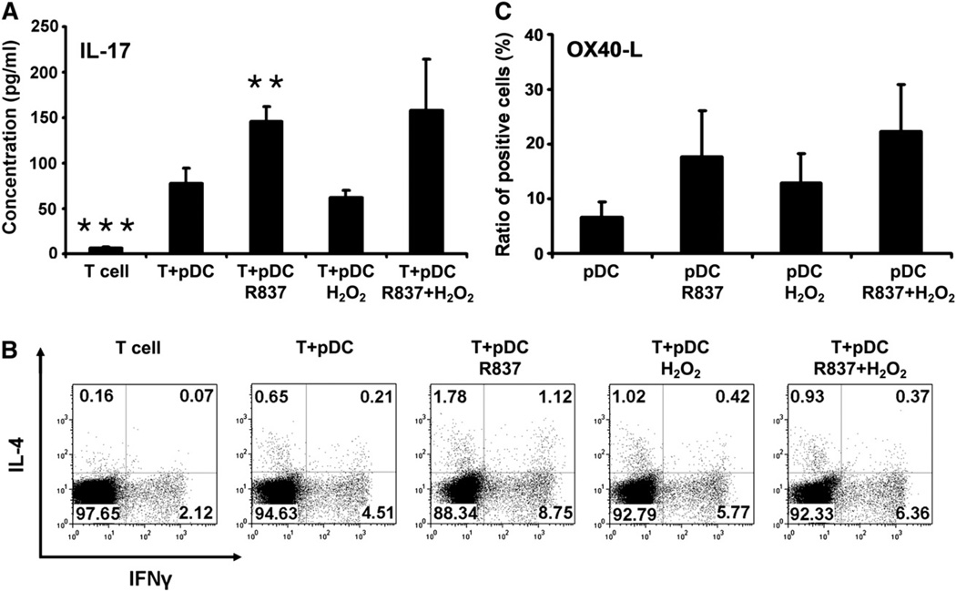
Fig. 6.
Th-polarizing ability of pDCs exposed to H2O2. Freshly isolated pDCs were treated with 0.01 µM H2O2 and R837, separately and in combination for 24 h, and then washed and cocultured for up to 6 days with naïve autologous CD4+CD45RA+ T cells. (A) Th17-polarizing ability of H2O2-treated pDCs. To assess the development of Th17 cells in the cocultures, levels of secreted IL-17 were determined in the culture supernatants by ELISA. (B) Th1- and Th2-polarizing ability of H2O2-exposed pDCs. After 6 days of cocultivation, T cells were restimulated with anti-CD3 mAb, phorbol 12-myristate 13-acetate, and ionomycin, and intracellular IFN-γ and IL-4 staining was performed. The percentages of IFN-γ- and IL4-positive cells were analyzed by means of flow cytometry. The dot-plot diagrams represent the results from one of four individual experiments. (C) Expression of OX40-L, a regulator of Th2 cell development, on the surface of pDCs treated with H2O2. Freshly isolated pDCs were stimulated with H2O2 only, R837 only, or both together for 24 h and then stained for OX40-L. Changes in the frequency of OX40-L-positive cells were analyzed using flow cytometry. Results are presented as means ± SE of three individual experiments. **P<0.01, ***P<0.001 vs T cells cocultured with untreated pDCs.

