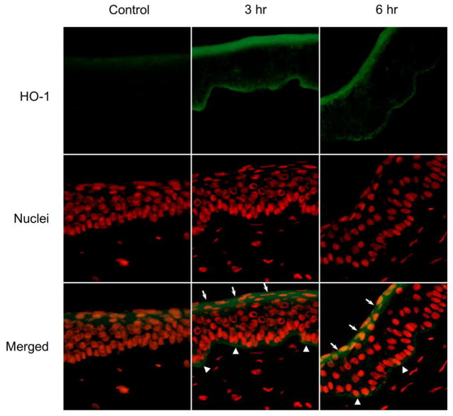
Figure 4. Effects of UVB on HO-1 expression in rabbit corneas.
Cornea organ cultures were exposed to control or UVB (0.5 J/cm2). After 3 hr and 6 hr, histological sections were prepared and the central portions of corneas analyzed for HO-1 expression using mouse monoclonal primary HO-1 antibody and Alexa-Flour 488-labeled secondary antibody. Nuclei were visualized using DAPI staining. White arrows and arrowheads indicate areas of HO-1 formation on the apical epithelial surface and basal epithelial surface, respectively. Original magnification x 400

