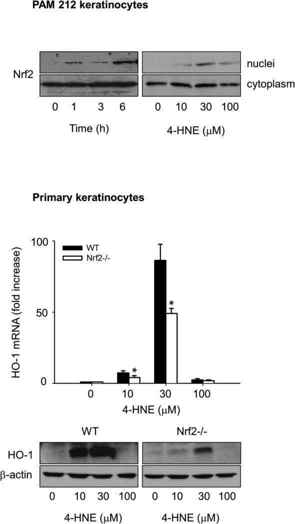
Figure 5. Role of Nrf2 in 4-HNE-induced HO-1 expression in mouse keratinocytes.
Keratinocytes were incubated with control or 4-HNE and analyzed for Nrf2 or HO-1 expression. Upper panel. Effects of 4-HNE on nuclear localization of Nrf2. PAM 212 cells were treated with 30 μM 4-HNE for 0, 1, 3 and 6 h or 0, 10, 30 and 100 μM 4-HNE for 3 h. Nuclear and cytoplasmic fractions of the cells were then prepared and Nrf2 expression analyzed by western blotting. Lower panel. Effects of Nrf2 expression on 4-HNE-induced HO-1 expression. Primary keratinocytes from wild type and Nrf2−/− mice were treated with 0, 10, 30 and 100 μM 4-HNE for 6 h and expression of HO-1 mRNA (upper panel) and protein (lower panel) analyzed by real-time PCR and Western blotting, respectively, as described in the Materials and Methods. HO-1 mRNA expression is presented as fold-increase relative to control. Each bar represents the mean ± SE (n = 3).

