Figure 1.

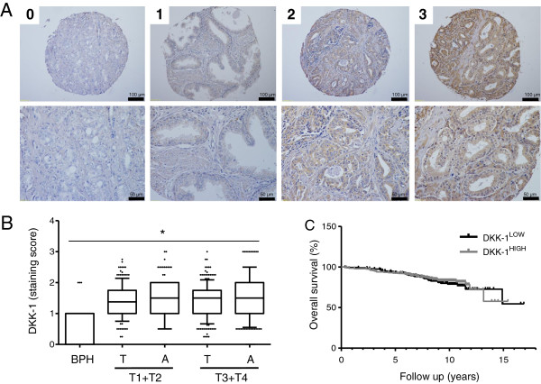
DKK-1 tissue expression in prostate cancer. A) The prostate TMA was immunohistochemically stained for DKK-1. Exemplary samples of each staining intensity (0–3) are shown. B) Distribution of DKK-1 expression in benign prostate hyperplasia (BPH), tumour tissue (T) and adjacent non-tumor tissue (A) is shown by boxplots. *DKK-1 tissue expression differed significantly between the different pT stages and the BPH tissues (p < 0.0001). C) Kaplan Meier survival analyses for PCa patients on the TMA dichotomized according to the median DKK-1 scores into high and low DKK-1 expression revealed no significant differences in overall survival (log-rank test: p = 0.27).
