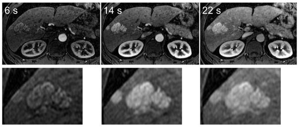
FIG 3.

FNH lesions can be seen with high resolution using the IVD method. Three out of five time-resolved images are shown for brevity. In this subject, images depicting two FNH lesions that were acquired at 6 seconds, 14 seconds, and 22 seconds after the start of the scan are shown. Magnified views depict the fine lobular edge features of the lesions, as well as the rapid enhancement pattern in these lesions.
