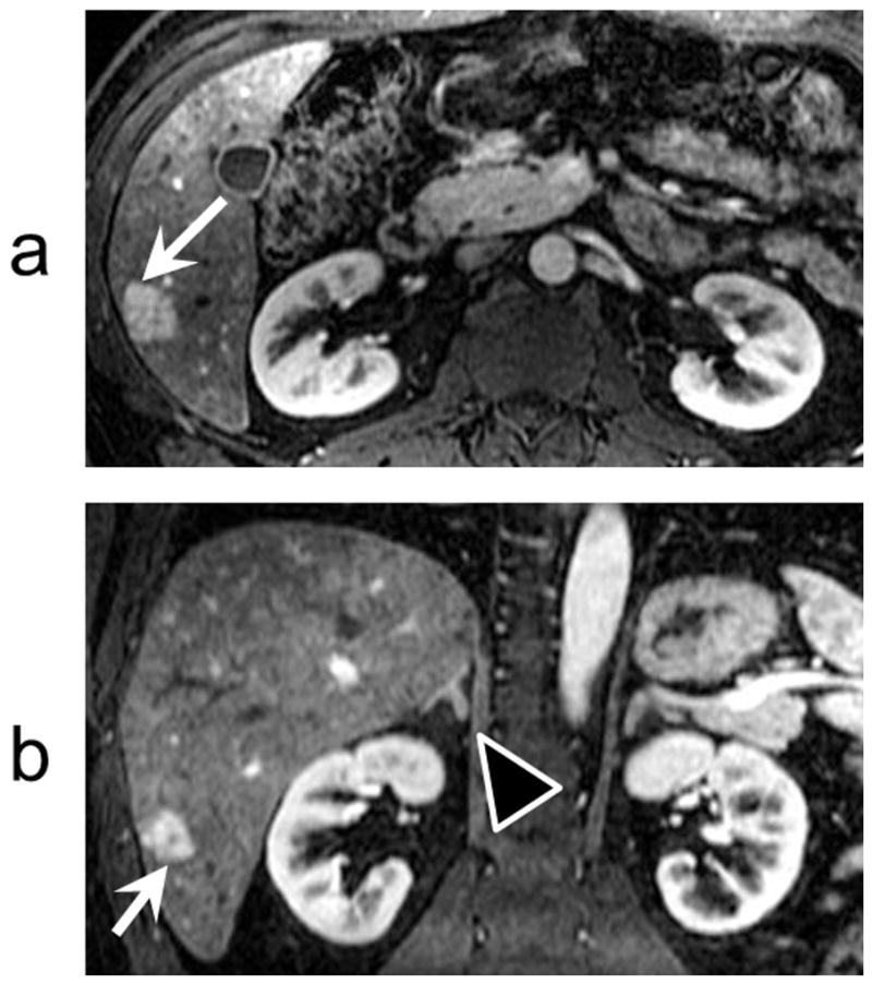
FIG 5.

IVD images were acquired with near-isotropic spatial resolution (1.2 mm × 1.7 mm × 2.6 mm). High spatial resolution enables assessment of the liver lesions in different planes. a) A cropped axial time-frame image from a subject with an FNH (white arrows). b) A cropped coronal reformat of the image in (a). Note the central scar in the FNH lesion in both the axial and coronal planes. Also note the excellent depiction of the right adrenal gland (black arrow head) and renal cortex. The full S/I coverage is 26 cm.
