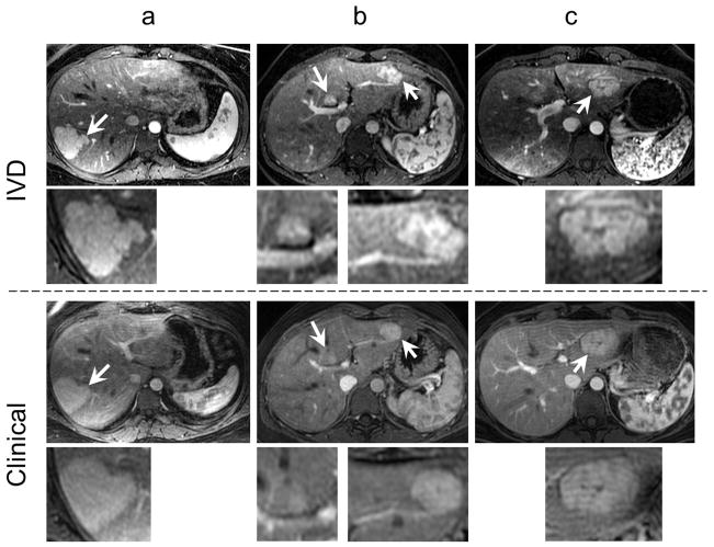
FIG 6.
The IVD images show superior quality compared to the clinical MR images. The top two rows show a late-arterial phase time-frame from the IVD series with magnified views of the lesions while the bottom two rows show the single late-arterial phase image from the clinical MRI protocol, in three different subjects with magnified views of the same lesions. Arrows point to the FNH lesions. Note the improved spatial resolution and lesion conspicuity in the IVD images relative to the clinical MR images.

