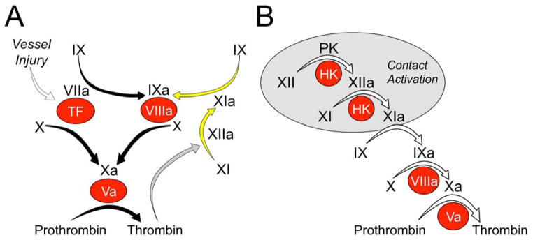
Figure 1.

Models of thrombin generation. (a) Tissue factor (TF)-initiated thrombin generation. Factor (f)VIIa binds to TF, a membrane protein expressed on the surface of cells underlying the blood vessel endothelium. The fVIIa–TF complex activates fX to fXa (the traditional extrinsic pathway of coagulation), and fIX to fIXa. FXa converts prothrombin to thrombin in the presence of fVa. fIXa sustains the process by activating additional fX in the presence of fVIIIa. The reactions indicated by the black arrows form the core of the thrombin generation mechanism in vertebrate animals. Mammals have fXIa, which provides another mechanism for fIX activation. In the traditional intrinsic pathway of coagulation fXIIa converts fXI to fXIa. fXI can also be activated by thrombin generated early in the coagulation process (gray arrow), explaining the lack of a bleeding disorder in people lacking fXII. (b) Contact-activation-initiated thrombin generation. In the cascade or waterfall model of thrombin generation, fXII is converted to fXIIa by a process called contact activation (gray circle) that requires prekallikrein (PK), high molecular weight kininogen (HK) and a negatively charged surface. fXIIa then activates fXI, setting off the sequence of proteolytic reactions that culminates in thrombin generation. In both panels zymogens of trypsin-like enzymes are indicated in black lettering, with active forms indicated by a lower case ‘a’. Non-enzyme cofactors are indicated by red circles.
