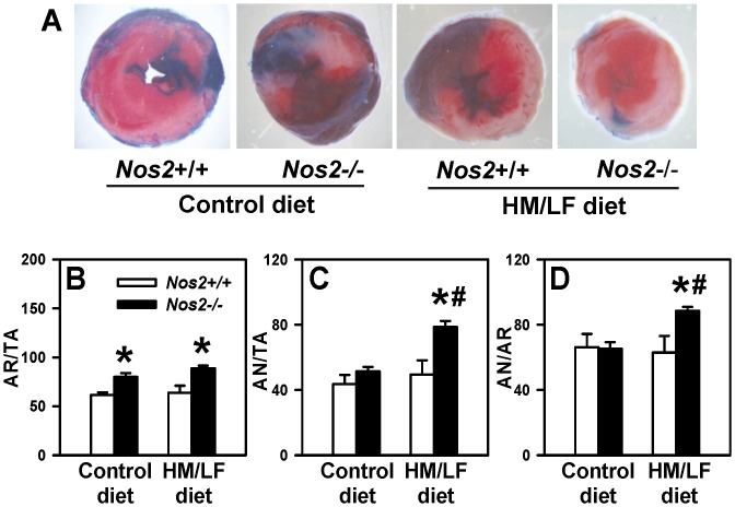
Figure 5. Regional myocardial ischemia-reperfusion injury.
The left coronary artery was ligated for 30 minutes and then reperfused for 2 hours. Panel A illustrates representative 2,3,5-triphenyltetrazolium chloride (TTC) stained images for each of the four groups of mice. The percent of (B) area at risk over total area (AR/TA), (C) area of necrosis over total area (AN/TA), and (D) area of necrosis over area at risk (AN/AR) were calculated in Nos2+/+ (open bars) or Nos2−/− mice (filled bars) by staining with Evans blue and TTC. Values are mean ±SE. 8–12 mice were studied in each group. *P<0.01 compared with Nos2+/+ mice fed the same diet, #P<0.05 compared with mice of the same genotype fed the control diet by two-way ANOVA.

