Figure 1.

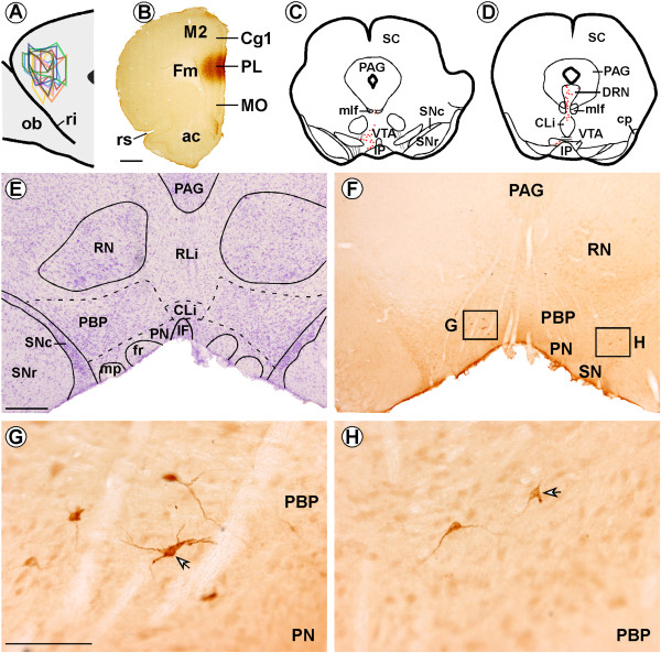
Location of Fluorogold (FG) injections in the prelimbic region (PL) of the medial prefrontal cortex and FG-labeled neurons in the ventral tegmental area (VTA). (A) Sagittal scheme shows all FG infusions in PL (n = 10; modified from Swanson, 1998). (B) Coronal section showing an FG deposit in PL using immunohistochemistry in one animal. (C-D) Representative coronal brainstem drawings showing FG-immunolabeled neurons (red dots) in the VTA of one of the PL group rats. (E) Nissl-stained section adjacent to the section shown in F, which delineates the subdivisions of VTA. (F) Panoramic photomicrograph showing FG-labeled neurons in the parabrachial subdivision (PBP) of VTA. (G) High magnification of box “G” from F showing FG-labeled neurons in the PBP of VTA ipsilateral to the FG injection site. The peroxidase immunoreaction product can be clearly observed in the cytoplasm of cell bodies and proximal dendritic branches (arrow). (H) High magnification of box “H” from F, showing much weaker FG-retrograde labeling (arrow) in the PBP of VTA contralateral to the FG injection. ac, anterior commissure; Cg1, cingular cortex, CLi, caudal linear raphe nucleus; DRN, dorsal raphe nucleus; Fm, forceps minor of the corpus callosum; fr, fasciculus retroflexus; IF, interfascicular subdivision of VTA; IP, interpeduncular nucleus; M2, secondary frontal cortex; mlf, medial longitudinal fasciculus; MO, medial orbital cortex; ob, olfactory bulb; mp, mammillary peduncle; PAG, periaqueductal gray; PN, paranigral subdivision of VTA; ri, rhinal incisure; RLi, rostral linear raphe nucleus; RN, red nucleus; rs, rhinal sulcus; SC, superior colliculus; SNc, substantia nigra pars compacta; SNr, substantia nigra pars reticulata;. Scale bars, B, 1 mm, E-F, 500 μm, G-H, 100 μm.
