
Figure 6.
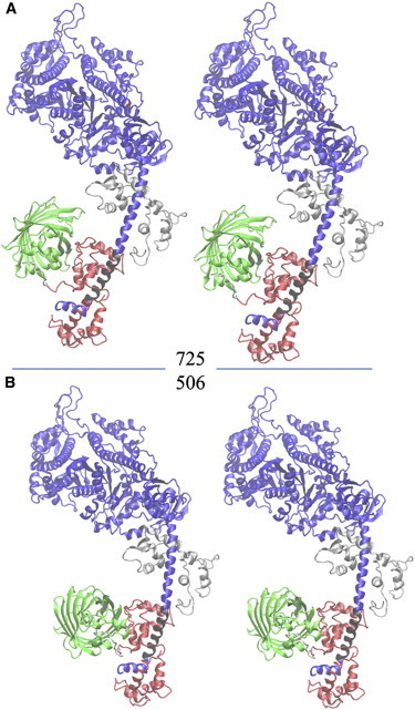
Stereo representations of S1/GFP. The myosin sequence shows the MHC (blue or black), the RLC (red), the GFP (green), and the ELC (silver). The 11-residue sequence linking the RLC C terminus to the GFP N terminus is white. The black section of the MHC where RLC binds to the lever arm defines an α-helix symmetry axis with spherical polar coordinates (β,α) defined relative to the lab coordinates (Lx′,Ly′,Lz′) in Fig. 2. (A) Structure 725 is the best S1/GFP representation of fish skeletal myosin. (B) Structure 506 is the best S1/GFP representation of porcine cardiac myosin.