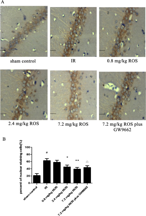
Figure 3.

Effects of rosiglitazone on NF-κB expression in hippocampal tissues of rats with global cerebral IRI (HE stain, ×400, n = 3). A: representative images of the hippocampal CA1 region after IRI. (Scale bars = 50 μm). B: Group data showing the effect of rosiglitazone on NFκB expression and translocation. #P < 0.05 compared with sham control group; *P < 0.05 **P < 0.01 compared with the IR group; ΔP < 0.05 compared with 7.2 mg/kg ROS group.
