
Fig. 1.
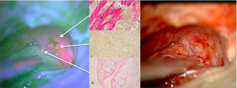
Left: Red fluorescent atypical meningioma after 5-ALA with fluorescence in tumor, adjacent dura and arachnoid overlying cortex. Right: Corresponding white light image. Center: Elastica-van-Gieson staining of biopsies. Top: Adjacent, fluorescing dura (“d”); middle: gross tumor and bottom: invaded brain (“c”: cortex)