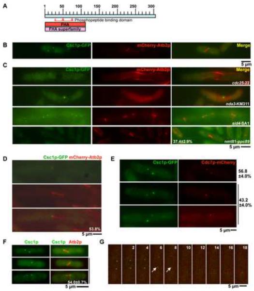
Figure 1. Csc1p, a new Forkhead-associated domain containing protein localizes to both SPBs and then transiently to one SPB during early mitosis.
A. Schematic representation of Csc1p with the FHA domain at the N-terminus. The phosphopeptide binding domain is highlighted between amino acids 30 and 80. B. Csc1p localizes to both SPBs in cells with short mitotic spindles. csc1-GFP mCherry-atb2 cells were grown at 24°C till mid-log phase, fixed with methanol and the localization of Csc1p imaged. C. Csc1p localization depends on entry into mitosis and the spindle pole body component Ppc89p, but does not require microtubules and SIN activity. cdc25-22 cells and sid4-SA1 cells expressing Csc1p-GFP and mCherry-Atb2p were grown at 24°C, shifted to the semi-permissive temperature of 33°C for 5 hours and imaged for the two fluorescent proteins. nda3-KM311 cells expressing Csc1p-GFP and mCherry-Atb2p were grown at 30°C, shifted to the non-permissive temperature of 19°C and imaged for the two fluorescent proteins. nmt81-ppc89 cells expressing Csc1p-GFP and mCherry-Atb2p were grown in EMM medium containing thiamine for 36 hours. The cells were then fixed with methanol imaged and for the two fluorescent proteins.(n=50×3) D. Csc1p shows a transient asymmetric localization to one SPB. cdc25-22 cells expressing Csc1p-GFP and mCherry-Atb2p were arrested at G2 by incubation at 33°C for 3 hours. The cells were then released at 24°C and grown for 1 hour. Cells were fixed with methanol and the localization of Csc1p and microtubules imaged. The percentage of cells with asymmetric distribution of Csc1p is shown at the bottom of the figure (n=26). E–G. Csc1p and Cdc7p segregate to opposite SPBs. E. cdc25-22 cells expressing Csc1p-GFP and Cdc7p-mCherry were grown at the permissive temperature and fixed with methanol. The fluorescence patterns of Csc1p and Cdc7p were scored. Cells that showed signals of both Csc1p and Cdc7p were imaged to analyze the localization relationship of the two proteins at SPB(s). The percentage of cells showing symmetric distribution (top cell) and asymmetric distribution (bottom two cells) of Cdc7p and Csc1p are indicated (n=50×3). F. Csc1p-GFP asymmetry is observed in wild-type cells. Wild-type cells expressing Csc1p-GFP and mCherry-Atb2p were grown at 24°C till mid-log phase, fixed with methanol and scored for the symmetric or asymmetric distribution of Csc1p in cells with short spindles (n=50×3). G. cdc25-22 cells expressing Csc1p-GFP and Cdc7p-mCherry were grown at the permissive temperature and imaged by time-lapse microscopy. Elapsed time (in minutes) after start of the imaging is indicated above the panels. The arrows indicate positions when asymmetry of Cdc7p and Csc1p are clearly apparent.

