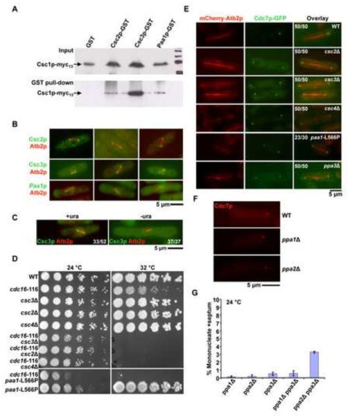
Figure 3. Identification of a multiprotein complex (SIP) required for generation of Cdc7p asymmetry.
A. Csc2p, Csc3p, and Paa1p bind Csc1p-Myc13. GST, Csc3-GST, Csc2-GST and Paa1-GST were isolated from yeast cells expressing Csc1p and one of these proteins using glutathione linked agarose beads. The isolated proteins were immunoblotted with an antibody against the Myc-epitope. The top panel shows the total lysates probed with the anti-Myc antibody, whereas the bottom panel shows an immunoblot of the proteins isolated from glutathione-agarose beads and probed with the anti-Myc antibody. B. Csc2p, Csc3p, and Paa1p localize to early mitotic SPBs. Cells individually mCherry-Atb2p and one of the following fusion proteins, Csc2p-GFP, Csc3p-GFP and Paa1p-GFP were grown at 24°C and the localization patterns of the fluorescent proteins investigated. Shown are 3 examples in each case, of cells with short mitotic spindles. C. The SPB component Ppc89p is required for Csc3p localization to SPB. Spores from a ppc89::ura4+/ppc89+ strain expressing Csc3p-GFP were germinated in minimal media in the presence or absence of uracil. The cells were then observed for the localization of Csc3p during early mitosis. The proportion of spores germinated in medium containing uracil and exhibiting and exhibiting a short spindle and Csc3p on SPBs is indicated in the figure. In addition, the proportion of spores germinated in the absence of uracil and exhibiting a short spindle and Csc3p on the SPB is indicated in the figure. D. csc2Δ, csc3Δ, csc4Δ, and paa1-L566P display synthetic lethal interactions with cdc16-116. Serial dilutions of csc2Δ cdc16-116, csc3Δ cdc16-116, csc4Δ cdc16-116 and paa1-L566P cdc16-116 and the corresponding single mutants were spotted on YES-agar medium and incubated at 24°C or at 30°C. E. Csc2p, Csc3p, Csc4p, Paa1p, and Ppa3p are required for the generation / maintenance of Cdc7p asymmetry. csc2Δ, csc3Δ, csc4Δ, and ppa3Δ cells expressing Cdc7p-GFP and mCherry-Atb2p were grown at 24°C, fixed with methanol and imaged for Cdc7p localization during late anaphase. paa1-L566P cells expressing Cdc7p-GFP and mCherry-Atb2p were grown at 24°C and then shifted to 36°C and the localization of Cdc7p during late anaphase investigated. The proportion of cells exhibiting symmetric localization of Cdc7p in the various mutants and the proportion of wild-type cells showing asymmetric distribution of Cdc7p are indicated. F. Cdc7p asymmetry is not compromised in the absence of Ppa1p and Ppa2p. Wild-type, ppa1Δ, and ppa2Δ cells expressing mCherry-Cdc7p were grown at 24°C and the localization of Cdc7p during late anaphase investigated. G. Combined loss of Ppa2p and Ppa3p leads to a modest increase in uninucleate cells with division septum. ppa1Δ, ppa2Δ, ppa3Δ, ppa1Δ ppa3Δ, and ppa2Δ ppa3Δ cells were cultured in the presence of 12 mM hydroxyurea and the proportion of uninucleate cells with a division septum estimated (n=500×3).

