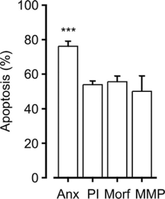
Figure 2.

Comparison of percentages of spontaneous eosinophil apoptosis obtained by different apoptosis determination methods. Apoptosis was determined by Annexin-V FITC/propidium iodide double-staining (Anx, n = 12), DNA fragmentation assay carried out by propidium iodide staining of permeabilized eosinophils (PI, n = 56), morphological analysis of May-Grunwald–Giemsa-stained eosinophils (Morf, n = 23), or determination of mitochondrial membrane potential by JC-1 staining (MMP, n = 5) after 40 h of incubation. Descriptions of the methods used can be found at Ref. 73.
Note: ***Indicates P < 0.001 as compared to all other columns by using ANOVA analysis.
