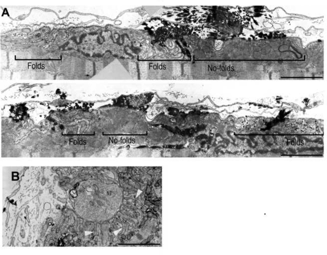
Figure 4.
Comparison of ultrastructure of agrin-induced, ectopic postsynaptic membrane and original neuromuscular junction in muscle fiber of soleus muscle. A) Montage of images taken along a longitudinal section of agrin-induced postsynaptic cluster. B) Electron microscopy of an innervated NMJ. Note the scattered distribution of synaptic folds along the ectopic postsynaptic membrane compared to the original synapse where folds are denser and deeper (see arrowheads). Black electrondense material in (A) marks reaction product from acetylcholine esterase activity used to locate the site of the ectopic AChR cluster. NT: nerve terminal. Bars, 3 µm

