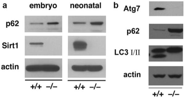
Fig. 5.

Accumulation of p62 as a marker of autophagic flux. (a) Accumulation of the autophagy marker p62 in the myocardium of embryonic and neonatal Sirt1−/− tissue. Neonatal tissue was harvested in the first 3 h after birth (reprinted with permission from Lee et al. [6]). (b) Analysis of wild-type and Atg7- deficient MEFs. Levels of p62 are increased and levels of LC3-II are decreased in Atg7−/− MEFs. These two conditions allow one to conclude that there is a defect in autophagy
