
Fig. 6.
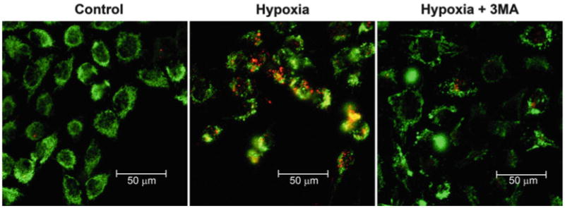
Detection of mitophagy using mtKeima fluorescence. Hela cells expressing mtKeima were exposed to hypoxic conditions for 24 h with or without the addition of 5 mM of 3-methyladenine (3MA). In this confocal micrograph, mtKeima fl uorescence signal from 561 nm (acidic) is red and the signal from 458 nm (neutral) is green. When mitophagy is induced, red punctate structures representing mitochondria contained within the acidic lysosome appear. The appearance of these red punctate structures can be inhibited by 3MA treatment