
Figure 2. Organotypic hippocampal slice cultures spontaneously develop epileptiform discharges in the growth medium.
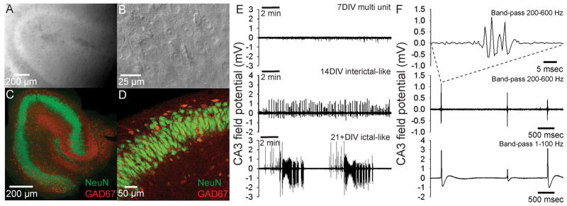
A. Low power IR-DIC image of hippocampal slice culture in the recording chamber shows preserved slice integrity and organotypic organization. B. High power IR-DIC image showing healthy pyramidal neurons in CA3. C. Immunohistochemical staining of a 30DIV slice with antibodies against NeuN (green) and GAD67 (red), respectively labeling neuronal nuclei and GABAergic cell bodies and processes. D. Higher power image showing CA3 pyramidal neurons labeled in green, GABAergic processes in red and double-labeled GABAergic interneuron cell-bodies in orange. E. Field potential recordings of spontaneous activity from CA3 of organotypic slice cultures. At 7 DIV only physiological multi-unit activity was recorded, but after 14 DIV the majority of slices displayed epileptiform, interictal-like spikes. This spontaneous epileptiform activity developed into a mix of interictal- and ictal-like discharges in slice cultures older than 21 DIV. F. Band-pass filtered (8-pole Bessel filter at 200 Hz, Gaussian filter at 600 Hz) field potential recording from area CA3 of a 14 DIV organotypic slice culture reveals fast ripple-like complexes (top trace). These fast ripple-like complexes occured at intervals larger than 1 sec (middle trace). Fast ripple-like complexes were associated with interictal-like spikes (bottom trace filtered w. 8 pole Bessel filter at 1 Hz, Gaussian filter at 100 Hz).