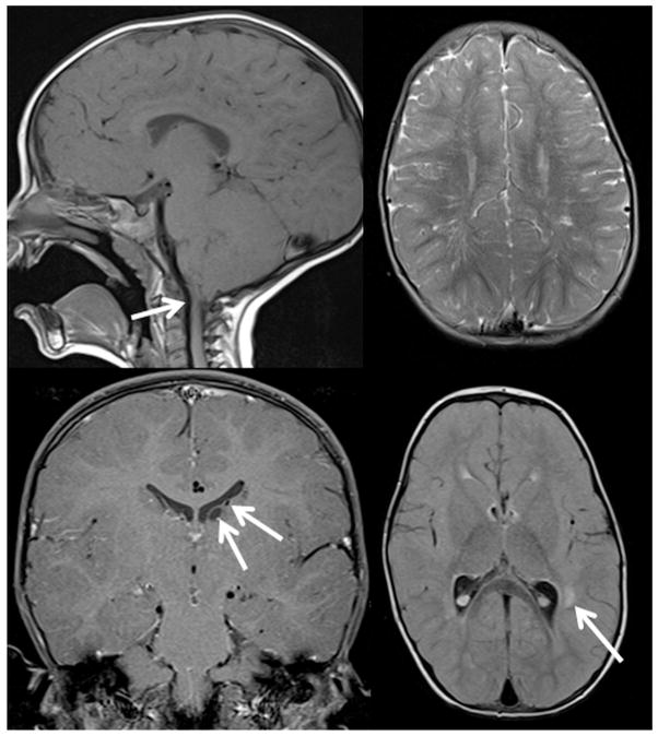
FIGURE 4.
Brain magnetic resonance imaging findings in a 26-month-old patient with bilateral uveal colobomas involving the iris and retina. (Top left) Type I Chiari malformation; the tips of the cerebellar tonsils lie 6 mm below the inferior margin of the foramen magnum (arrow), at the same level as the posterior arch of C1. (Top right) Prominent perivascular (Virchow-Robin) spaces are unusually numerous in the white matter of the cerebral hemispheres. (Bottom left) There are 2 subependymal cysts on the left side (arrow). (Bottom right) There are several nonenhancing lesions of indeterminate etiology (arrow) located in the perivascular white matter of the left temporal lobe.

