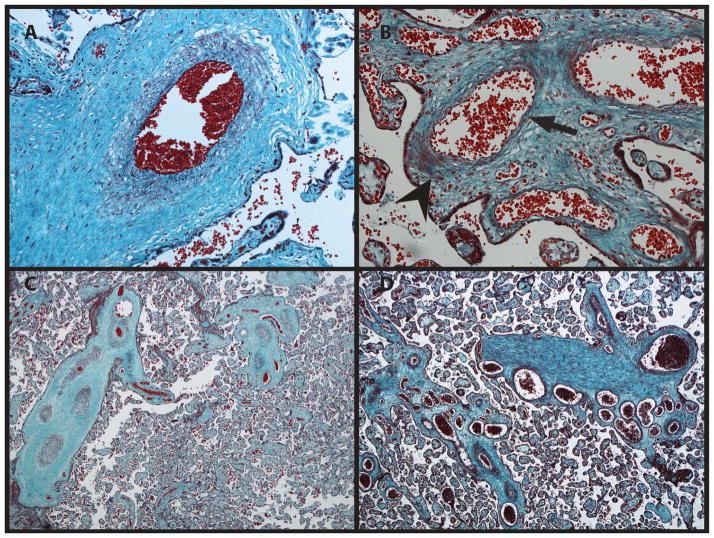
FIG. 1.
A. Microscopic image of a normal placenta at 200X stained with Masson trichrome. B. The patient’s placenta at 200X. The smooth muscular layer is markedly abnormal with an asymmetric layer of myocytes (arrowhead: clumped myoctyes; arrow: poor myocyte coverage), in keeping with failed pericyte migration. C. Control placenta at 40X. D. The patient’s placenta at 40X. The vessels are ectatic. A cluster of numerous veins is in keeping with tortuosity.

