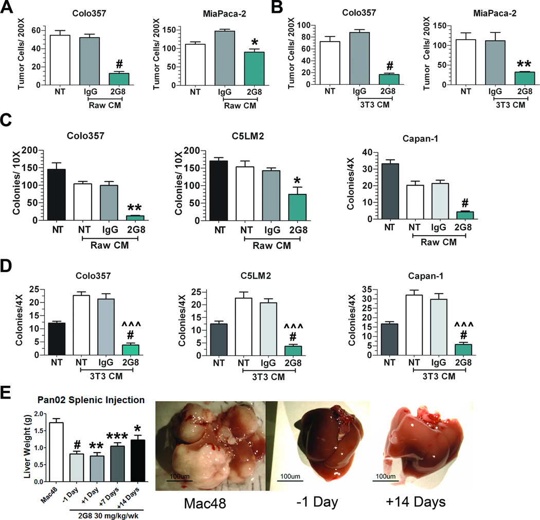
Figure 2. Inhibition of Tgfβr2 on stromal cells limits tumor cell migration and colony formation in vitro and in vivo.
A and B, Transwell migration of tumor cells (Colo357 and MiaPaCa-2) towards RAW 264.7 (A) or NIH 3T3 (B) cells treated with serum free media (NT), control Rat IgG (IgG) or 2G8 for 24 hours. After removing treatment conditions, Colo357 and MiaPaCa-2 cells were plated in transwell chambers and allowed to migrate overnight towards previously treated stromal cells. Bar graphs represent number of cells/200× field. The assays were performed in triplicate in two independent experiments. *, p<0.05; **, p<0.01; #, p<0.0001 vs. Rat IgG. C and D, Anchorage independent growth of tumor cells (Colo357, C5LM2, and Capan-1) in the presence of growth media with 10% serum (NT), conditioned media from RAW 264.7 (C) or NIH 3T3 (D) cells grown in media with 10% serum with or without control Rat IgG (IgG) or 2G8. Colony formation was quantified by number of colonies per well at 4× or 10× magnification. Bar graph represents mean +/− SEM of a single experiment performed in triplicate with similar results found upon 2 independent experiments. *, p<0.05; **, p<0.01; **, p<0.01; #, p<0.0001 vs. NT; ^^^, p<0.0001 vs Rat IgG. E, Pan02 cells were injected intrasplenically and liver weight used as a surrogate for tumor burden in the liver. Mice were treated with a control rat IgG (Mac48, n=14) or 2G8 initiated at -1 (n=10), +1 (n=3), +7 (n=8) or +14 (n=7) days post splenic injection. Representative images of livers from mice are shown. Results are expressed as mean+/−SEM. *, p<0.05; **, p<0.01; ***, p<0.001; #, p<0.0001 vs. control

