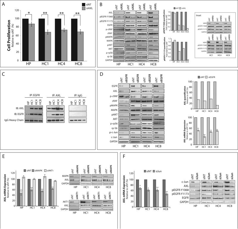
Figure 2. Cetuximab resistant cells depend on AXL and its cooperation with EGFR.
(A) Cells were transfected with AXL siRNA (siAXL) or non-targeting siRNA (siNT) for 72 hr prior to performing proliferation assays. Proliferation is plotted as percentage of growth relative to NT transfected cells (n=6 in 3 independent experiments). (B) Cells were incubated with AXL siRNA or NT siRNA for 72 hr prior to harvesting whole cell lysate and immunoblotting for the indicated proteins. GAPDH was used as a loading control. Phosphorylation of EGFR on tyrosine 1068 and 1173 were quantitated using Image J software. Inset: Cells were transfected with siRNA against HER2, HER3, or NT siRNA for 72 hr prior to harvesting whole cell lysate. GAPDH was used as a loading control. (C) 500 μg of whole cell lysate was subjected to immunoprecipitation analysis with cetuximab (IP:EGFR), anti-AXL (IP:AXL), or anti-IgG (IP:IgG) antibody followed by immunoblotting (IB) for either AXL or EGFR. IgG heavy chain staining from the IB:AXL blot was used as a loading control. (D, E, F) Whole cell lysate and mRNA were harvested from CtxR clones 72 hr post-transfection with EGFR siRNA (D), MAPK and AKT1 siRNAs (E), c-Jun siRNA (F), or NT siRNA. GAPDH was used as loading control for protein. In (D), AXL protein expression was quantitated using Image J software. AXL mRNA expression was detected by qPCR and normalized to AXL expression in siNT transfected cells (n=3 in 3 independent experiments). 18S was used as an endogenous control Data points are represented as mean± SEM. *p<0.05, **p<0.01.

