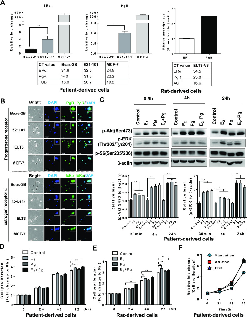
Fig. 1.

Progesterone activates Akt and ERK1/2 and enhances the proliferation of TSC2-deficient cells in vitro. a The relative transcript levels of ERα and PgR in patient-derived cells, rat-derived cells, MCF-7, and BEAS-2B cells. The actual CT values of the ERα and PgR transcript were shown in the tables. b Representative immunofluorescent staining images of ERα and PgR in patient-derived cells (621-101), rat-derived cells (ELT3-V3), BEAS-2B, and MCF-7 cells. c Patient-derived cells were treated with 10 nM E2, 10 nM progesterone (Pg), 10 nM E2 + 10 nM Pg, or vehicle control for 0.5, 4, and 24 h. Immunoblot analysis of phospho-Akt (Ser473), phospho-ERK1/2 (Thr202/Tyr204), and phospho-S6 (S235/236). A densitometry of phospho-ERK1/2 and ERK1/2 was performed. d LAM patient-derived cells were treated with 10 nM E2, 10 nM progesterone (Pg), 10 nM E2 + 10 nM Pg, or vehicle control for 24, 48, and 72 h. Cell proliferation was measured using crystal violet staining assay. e TSC2-deficient rat uterus-derived ELT3 cells were treated with 10 nM E2, 10 nM progesterone (Pg), 10 nM E2 + 10 nM Pg, or vehicle control for 24, 48, and 72 h. Cell proliferation was measured using crystal violet staining assay. f Patient-derived cells were in complete media (containing steroids) or in phenol red-free media supplemented with charcoal-dextran-stripped FBS (depleting steroids) for 72 h and measured cell growth at 24, 48, and 72 h post-cell seeding. Cell proliferation was measured using crystal violet staining assay. *p < 0.05, **p < 0.01, Student’s t test
