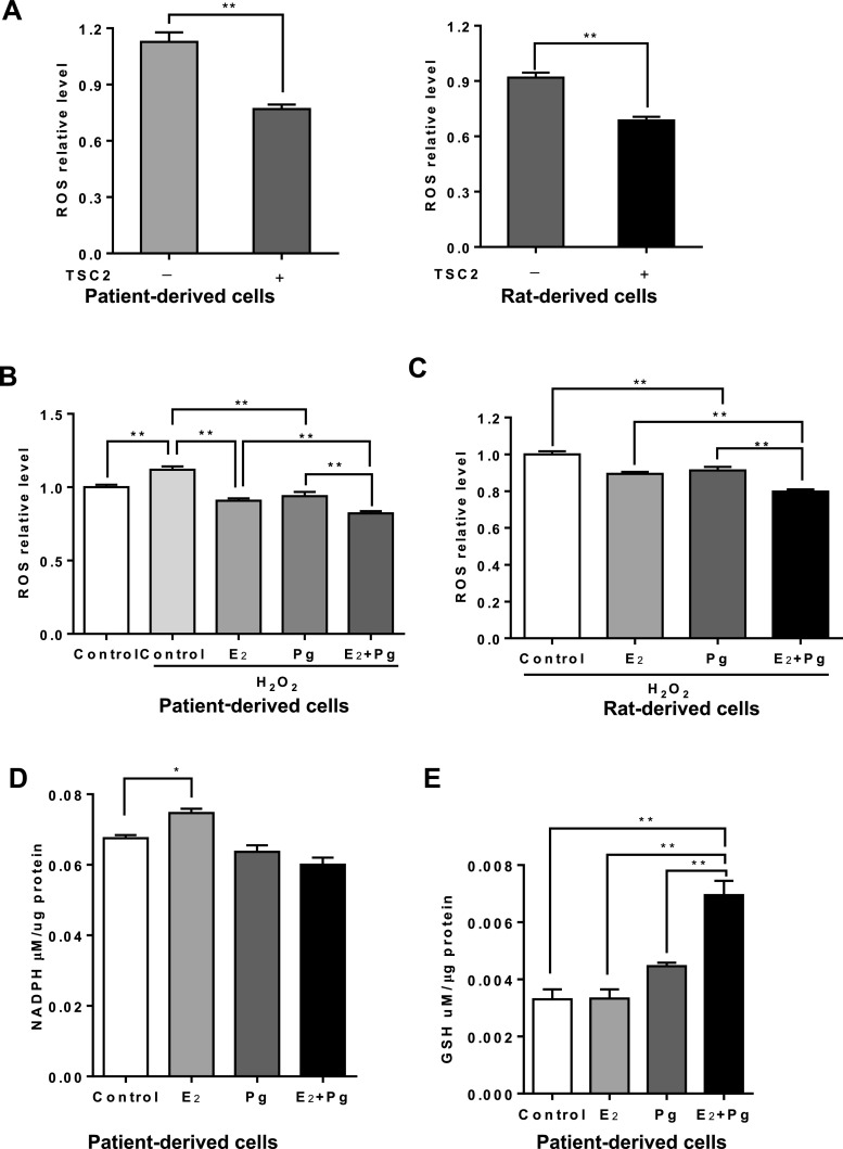
Fig. 4.
Progesterone and estradiol synergistically decrease ROS and increase GSH in TSC2-deficient cells. a Cellular levels of ROS were measured using DCFH-DA in patient-derived or rat-derived cells grown in serum-free conditions. b Rat-derived cells and c LAM patient-derived cells were treated with 10 nM E2, 10 nM Pg, 10 nM E2 + 10 nM Pg, or vehicle control for 24 h. Cells were incubated with hydrogen peroxide (0.5 μM) for 30 min prior to the measurement. Cellular levels of ROS were quantified using DCFH-DA. Data was normalized to total cell number. Results are representative of eight independent samples per group from three experiments. d Cellular levels of NADPH were measured in LAM patient-derived cells treated with 10 nM E2, 10 nM Pg, 10 nM E2 + 10 nM Pg, or vehicle control for 24 h. Data was normalized to total protein level. e Cellular levels of GSH were measured in patient-derived cells treated with 10 nM E2, 10 nM Pg, 10 nM E2 + 10 nM Pg, or vehicle control for 24 h. Data was normalized to total protein levels. Results are representative of three sets of independent samples per group from three experiments. *p < 0.05, **p < 0.01, Student’s t test

