
Figure 6. Recruitment of GFP-expressing M2 macrophages to SNpc in 6-OHDA-intoxicated mice.
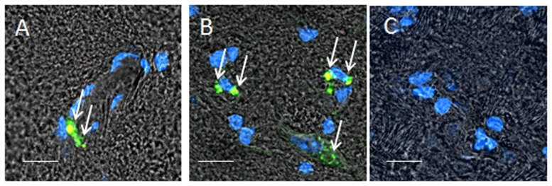
Macrophages were transfected with GFP-encoding pDNA as described in Materials and Methods section and stained with primary antibodies to CD206, a marker for M2 macrophages, and secondary fluorescently-labeled anti-Mouse-IgG-atto 647N (red). BALB/c mice were i.c. intoxicated with 6-OHDA into SNpc. Twenty one day later, the animals were i.v. injected with GFP-expressing RAW 264.7 macrophages (green, 5×10⁶ cells/ mouse in 100 µl). Twenty four hours later mice were sacrificed, and perfused with PBS and 4% PFA. Brains were frozen, sectioned with a cryostat (10 µm thick), and examined by confocal microscopy (60×magnification) (A, B). Healthy mice without brain inflammation (with PBS i.c. injections) were used as a control group (C). Slides were stained with for expression of mannose receptor (CD206 antibodies). Co-localization of GFP-expressing macrophages and CD206 antibodies to mannose receptor manifested in yellow staining (arrows) confirmed presence of significant amounts of the M2 genetically-modified cells in the intoxicated brain endothelial microvesseles (A), and parenchyma (B). No fluorescence in the healthy brain was found (C) indicating that systemically administered Raw 264.7 macrophages did not cross the BBB in the absence of brain inflammation. The bar: 20 µm.