
Fig. 6. Effects of knockdown of EGFR and β-catenin on U-87 MG cell migration and invasion.
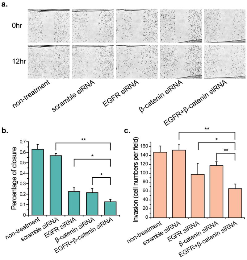
(a) Representative bright-field images of U-87 MG cell migration across the wound edges at 0–24 h after wounding in confluent U-87 MG transfected with scramble and targeted siRNA. (b) The bar graph represents the quantitative results for percent closure of cells migrating to the wound area. Analyses were performed at two randomly positions, and the error bars indicate the SD of the two independent analyses. (C) Decreased numbers of invading U-87 MG cells were observed after siRNA transfection in the transwell invasion assay. Asterisk indicates significant level in Student's t-test: *P < 0.05, **P < 0.01.