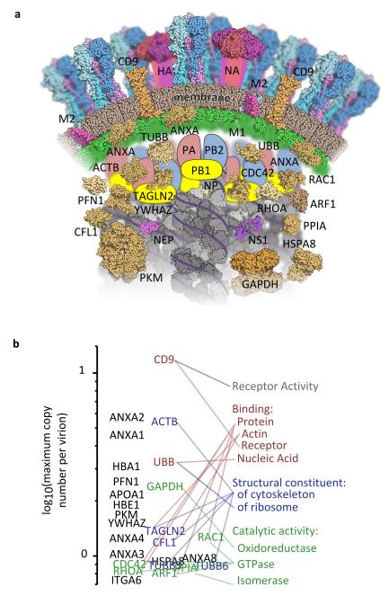
Figure 4. The architecture of an influenza virion.
Proteins in virions produced by WSN-infected bovine epithelial (MDBK) cells, present at more than one tenth the abundance of the viral polymerase and found in material purified both with and without HAd in at least two separate experiments. (a) Schematic cross-section of an influenza virion, showing proteins whose localisation in the virion is known or can be inferred from other studies. Host proteins and membrane are brown and viral proteins are brightly coloured. (b) Host proteins, ranked by maximum copy number in virions (calculated as in Fig. 2) and linked to their Gene Ontology molecular function terms.

