
Figure 5. Tethering Rhi suppresses EGFP expression and splicing.
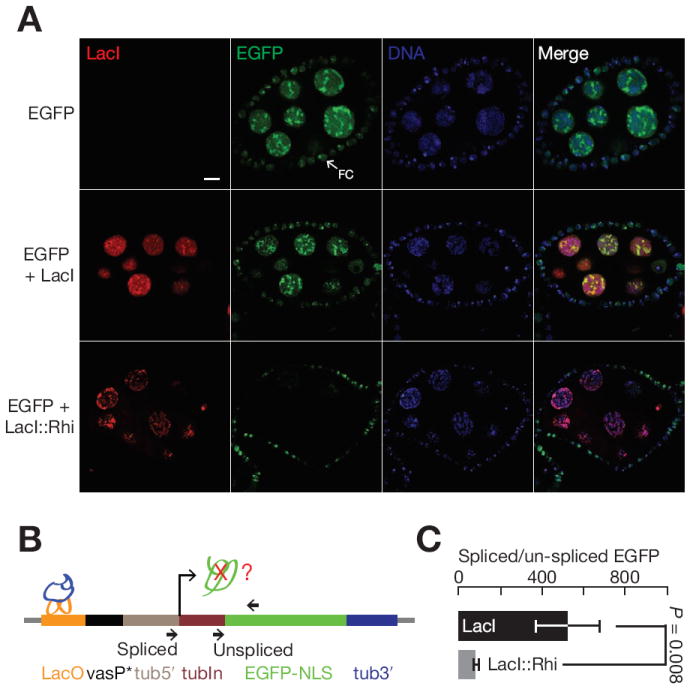
(A) Germline expression of LacI (red) does not suppress EGFP expression (green). By contrast, expression of LacI∷Rhi (red) suppresses EGFP accumulation in the nurse cell nuclei (green). Note that the EGFP reporter is expressed in both the germline and surrounding somatic follicle cells (arrows, FC). The fusion does not expressed in the follicle cells, and EGFP expression in these cells is not reduced. The bar in the up right panel is 10 μm, and applies to all panels.
(B) Splicing at the target locus. The diagram shows the target transgene and indicates that position of LacI or LacI∷Rhi binding (LacO) and the primers used to assay both spliced and unspliced transcripts by qRT-PCR.
(C) Bar graph showing the ratio of spliced to unspliced target in the presence of LacI (black) or LacI∷Rhi (grey). LacI∷Rhi binding lead to a significant reduction in splicing efficiency (p = 0.008), data are mean ± standard deviation for 3 independent biological samples.
See also Figure S6.