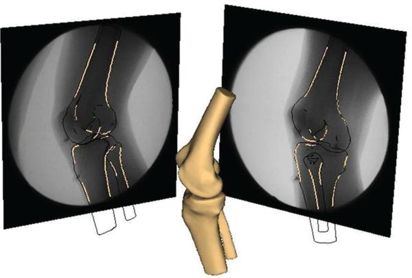
Figure 2.
An image of the reconstructed tibia/fibula and femur. Contours of bony landmarks were detected on each radiograph and a fully automatic 6 degrees of freedom optimization algorithm was used to determine the position and orientation that optimally matched the detected contours with the projected contours from the imported bone geometries.

