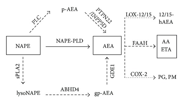
Figure 1.

Schematic illustration of parallel AEA synthesis and degradation pathways. NAPE-PLD represents a Ca2+-dependent route of AEA formation (—), while other enzymes (- - -) act in a Ca2+-insensitive manner. Main route of AEA degradation is hydrolysis by FAAH (- - -). High AEA tissue concentration triggers parallel catabolic pathways through LOX-12/15 and COX-2 enzymes (⋯).AEA: anandamide, NAPE: N-acylphosphatidylethanolamine, NAPE-PLD: N-acylphosphatidylethanolamine phospholipase D, GDE1: glycerophosphodiester phosphodiesterase 1, ABHD4: α/β hydrolase domain containing protein 4, PTPN22: protein tyrosine phosphatase nonreceptor type 22, sPLA2: soluble phospholipase A2, INPP5D: inositol 5-phosphatase, PLC: phospholipase C, FAAH: fatty acid amide hydrolase, COX-2: cycloxygenase 2, LOX-12: arachidonate 12-lipoxygenase, LOX-15: arachidonate 15-lipoxygenase, gp-AEA: glycerophosphoanandamide, p-AEA: phospho-anandamide, lysoNAPE: lyso-N-acylphosphatidylethanolamine, AA: arachidonic acid, ETA: ethanolamine, PG: prostaglandins, PM: prostamides, and 12/15-hAEA: 12/15-hydroxyanandamide.
