
Fig. 4.
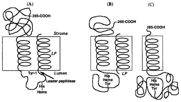
(A, B) Model for processing and assembly of intermediate forms of cyt f and (C) membrane topography of mature form. The schematic shows that complete heme ligation involving the α-amino group of Tyr-1 cannot occur until the 35-residue leader peptide (LP) has been removed by the processing peptidase that is believed to reside on the lumen side of the membrane (Johnson et al., 1991; Kirwin et al., 1991).