PLATE III.
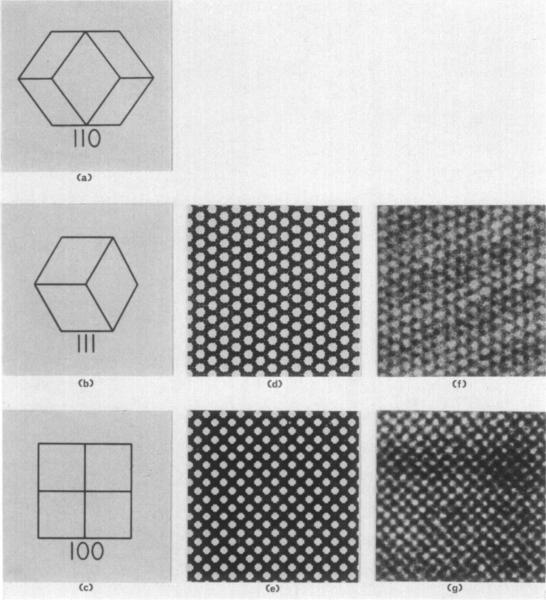
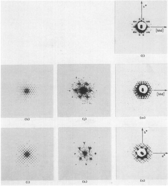
(a) to (c) Drawings of a rhombic dodecaherdral crystal viewed parallel to the 2, 3, and 4-fold axes, respectively. The crystal depicted in (a) is rotated 90° about the 2-fold axis with respect to the diffraction pattern to its right. The orientation of the crystal drawn in (b) is identical with the orientation of the sections and diffraction pattern3 in (d), (f), (h), (j), and (m). The crystal in (c) is rotated 45° with respect to the (e), (g), (i), (k), and (n).
(d) and (e) Projection drawings representing magnified (112,000 ×) images of the molecular packing in space group 14132 viewed perpendicular to the 111 and 100 crystal planes. Molecules are colored black to correspond with electron microgmphs of positively stained thin sections of embedded crystals appearing in (f) and (g). The drawings depict projections of the structure thicker than one unit cell.
(f) and (g) Electron micrographs of positively stained thin sections of fixed, dehydrated, and embedded form I RuDPCase crystals at a magnification of 112,000 × . Sections are perpendicular to the 3 and 4-fold axes of the crystal and are therefore views of the III and 100 crystallographic planes.
(h) and (i) Optica1 diffraction patterns of the drawings in (d) and (o) reproduced at the same scale as the X-ray precession photographs in (m) and (n).
(j) and (k) Optical diffraction patterns of the electron micrographs in (f) and (g). The patterns are enlarged four times larger than the scale of the diffraction patterns in (h) and (i) and (m) and (n).
(I) to (n) Three-degree zero-level precession photographs taken with the precession axis parallel to Lhe 2, 3, and 4-fold crystal axes. The central portion of each pattern is hidden hy the shadow of the lead beam stop . An image of the focused X-ray beam appears in the center of each pattern and defines t he position of the 0,0,0 reflection. The diffraction patterns have been magnified 4·5 times from the original photographs.


