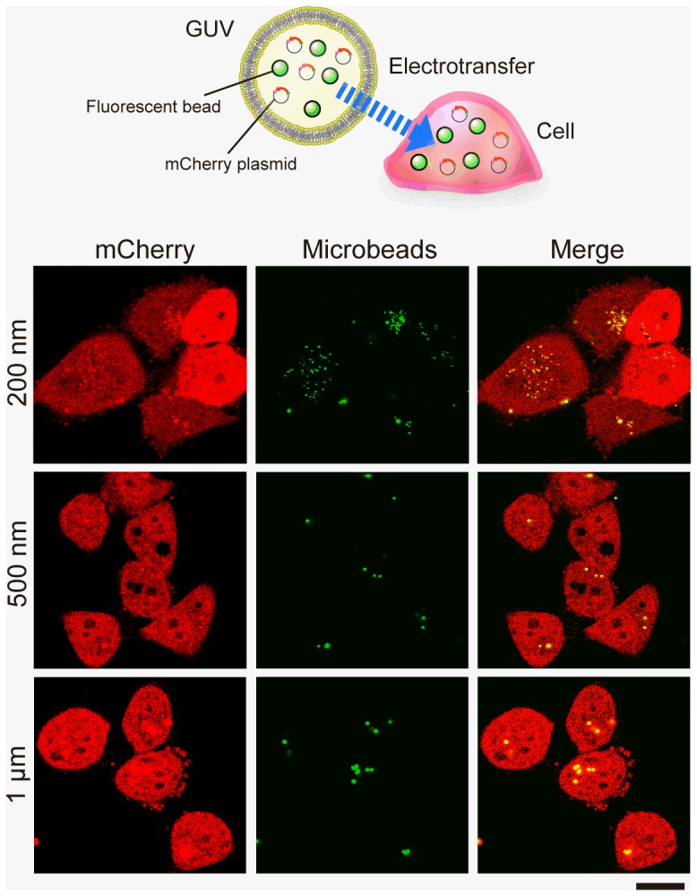
Figure 4. Introducing multiple components of microbeads and plasmids into live cells by cell–GUV electrofusion.

GUVs including both the plasmid mCherry and fluorescent microbeads were prepared for electrofusion with HeLa cells. The treated cells were then cultured for 2 days. Confocal microscopic images show the cross-section of the treated HeLa cells into which (from the top) beads of 0.2 µm, 0.5 µm, and 1 µm diameter (green) had been introduced. The mCherry expression in cells is shown in red, and merged images are shown in the right column. Scale bar = 20 µm.
