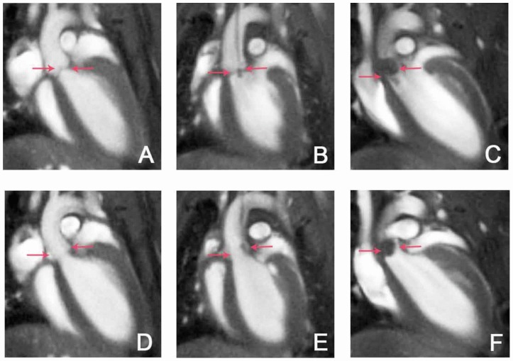
Figure 3. MRI detection of S. aureus vegetations on the aortic valve.
Exemplary long axis views of the mouse heart acquired with self-gated CINE UTE MRI (TR/TE : 5/0.31 ms, FA: 15°, resol: (125 µm)2, MTX: 256×256, slice thickness: 1 mm, scan duration: 12∶08 min) show the aortic valves in closed (A–C) and open (D–F) state. Flow artifacts were almost completely suppressed by the use of self-gated UTE MRI. (A,D) Images of a mouse with sham surgery infected with unlabeled bacteria show normal valves (arrows). (B, E) In images of a catheterized mouse infected with unlabeled bacteria the catheter as well as valve thickening and an additional intracardial mass is visible (arrows). (C, F) Large hypointensities on the valves (arrows) are observed in this catheterized mouse infected with iron labeled bacteria.

