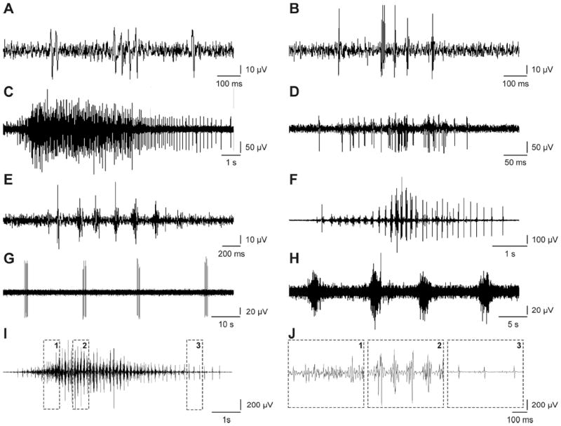
Fig. 2.

Examples of unit (A: BF data, 4-5pm; B: MG, 8-9am), tonic (C: BF, 2-3am; D: VL, 4-5am), clonus (E: MG, 6-7am; F: MG, 3-4pm), myoclonus (G: MG, 4-5am; H: MG, 7-8pm) and a mixed spasm (I: MG, 9-10am). (J) Regions within the mixed spasm are expanded to show tonic EMG (box 1), clonus (box 2) and unit potentials (box 3).
