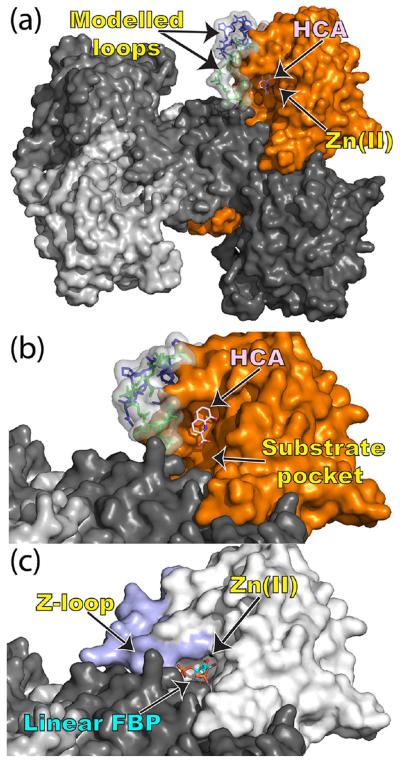
Figure 4.
Rearrangement of the substrate pocket caused by HCA binding. (a) Surface rendering of the MtFBA–HCA complex as its tetrameric biological assembly with one MtFBA protomer colored orange, the rest colored various shades of gray, and HCA rendered as pink sticks. Loops that were modeled in due to a lack of electron density are shown as transparent surfaces and either green sticks (residues 167–177) or blue sticks (residues 210–223). (b) Close-up of the substrate binding pocket of the MtFBA–HCA complex. (c) Close-up of the substrate binding pocket for MtFBA (white) bound with linear fructose 1,6-bisphosphate (cyan sticks; PDB entry 3ELF), in which residues 210–223 are colored light blue. In all cases, the zinc ions are rendered as a black sphere.

