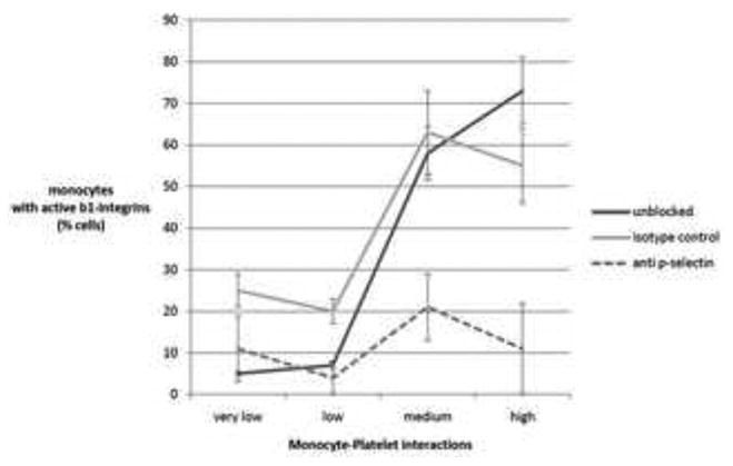
Figure 4. Effect of p-selectin exposure on monocyte integrins.

Plastic plates were coated with platelets at the relative density indicated by the x-axis. Platelets were then pretreated with anti p-selectin (dashed line) or an isotype control (gray line), or received no pretretment (black line). Monocytes were passed through the plates at 0.5 dyne/cm2 and then immediately assessed by flowcytometry for expression of active β1 integrins. The y-axis indicates the percent of monocyte which bound an antibody directed at an epitope only exposed when the integrin is in the open confirmation. Data shown are the means (+/- SD) of 3 independent experiments.
