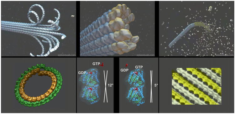
Fig. 1.

Structural view of microtubule dynamics, disassembly, and assembly intermediates, and nucleotide state of tubulin. Upper panels are schematics of three distinct states for microtubules: shrinkage (left), growth (right), and minimal guanosine triphosphate (GTP) cap (center). Lower panels are the experimental cryo-electron microscopy structures for stabilized mimics of the disassembly (left) and assembly intermediates (right), and the tubulin dimer structures for guanosine diphosphate-bound and GTP-bound tubulin outside the context of the microtubule lattice (center). Adapted from Figs. 1 and 2 in Nogales and Wang (2006). (See Plate no. 5 in the Color Plate Section.)
