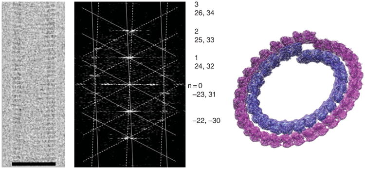
Fig. 3.

Double-layered guanosine diphosphate (GDP)-tubulin tubes. Cryo-electron microscopy image of a 32/24 double-layered GDP–tubulin tube (left) and its diffraction pattern (center). The pattern was indexed for the near and far side of the helix (solid and dashed lattice, respectively). The indexing indicates severe Bessel overlap of the two layers of helices on all the layer lines (Bessel orders for the inner and outer layer, respectively, are shown on the right). Docking of the tubulin crystal structure into one turn each of the inner and outer layer. Scale bar in left panel corresponds to 50 nm. (See Plate no. 7 in the Color Plate Section.)
