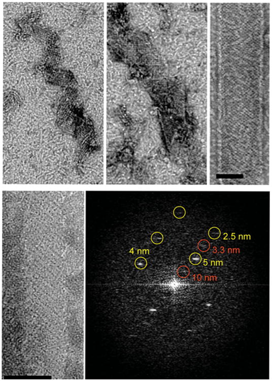
Fig. 4.

Growth of GMPCPP–tubulin ribbon. Time points in the growth of GMPCPP ribbons (top). Early assemblies already show the pairing of protofilaments as shown by the presence of strong 10 nm repeats perpendicular to the protofilament axis (bottom). Scale bars correspond to 50 nm.
