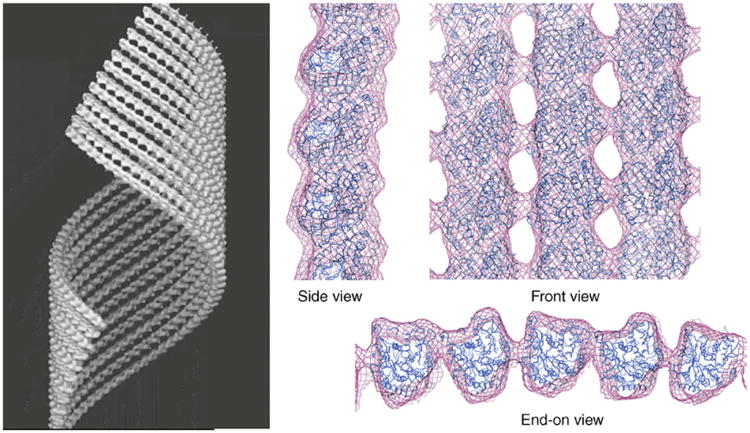
Fig. 5.

Structure of GMPCPP–tubulin tubes. Three-dimensional cryo-electron microscopy reconstruction where only 10 protofilaments of the tube wall are shown (left). Docking of the crystal structure of a tubulin monomer to reproduce the tubulin lattice in this structure, shown in different views (right). The side view shows the slight outward curvature of the GMPCPP protofilament. The front view shows the striking similarity of the lateral protofilament stagger in this structure and that of microtubules. In the end-on view the pairing of protofilaments is most apparent. Right side adapted from Fig. 3c in Wang and Nogales (2005). (See Plate no. 8 in the Color Plate Section.)
