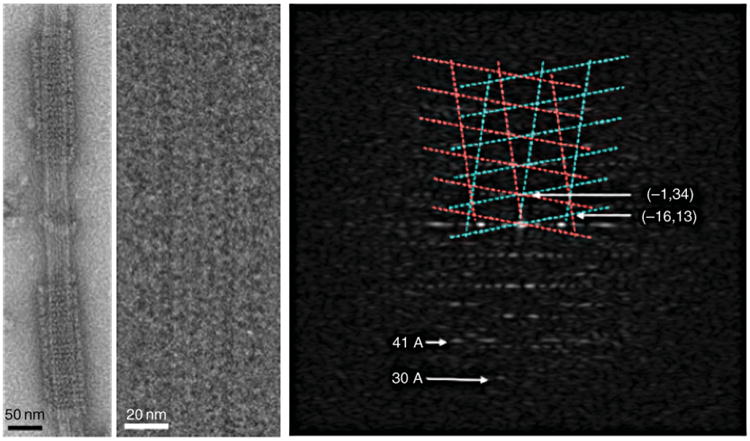
Fig. 6.

Self-assembly of Dam1 complexes into double spirals around microtubules. Negative stain image (left). Cryo-electron microscopy image of a small, ordered segment of spiral (center) and its diffraction pattern (right) extending to about 30 Å resolution. The Dam1 lattice is indicated in cyan and magenta. The tubulin 41 Å repeat is indicated. (See Plate no. 9 in the Color Plate Section.)
