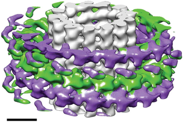
Fig. 7.

Cryo-electron microscopy reconstruction of the Dam1 double spiral around microtubules. The spirals run antiparallel (each shown in two distinct colors) and the resulting lack of polarity of the Dam1 assembly stands in contrast with the polar character of the underlying microtubule. Scale bar corresponds to 100 nm. (See Plate no. 10 in the Color Plate Section.)
