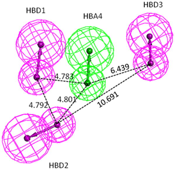
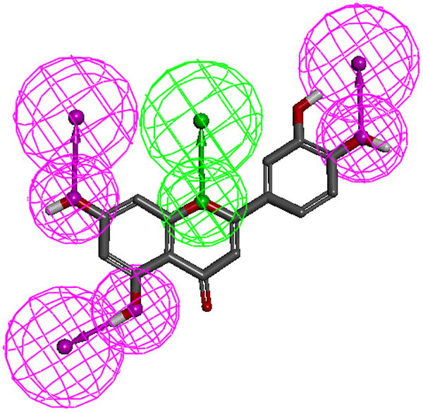
Figure 3.


A.The top-ranked pharmacophore model (1, see supplemental Table 1). Pharmacophore model-1 consists of three hydrogen bond donors (magenta spheres) and one hydrogen bond acceptor (green spheres). Inter-spatial distances are shown in Angstroms.
B. Luteolin mapped to pharmacophore model-1. Note only polar hydrogens are displayed on the molecules.
