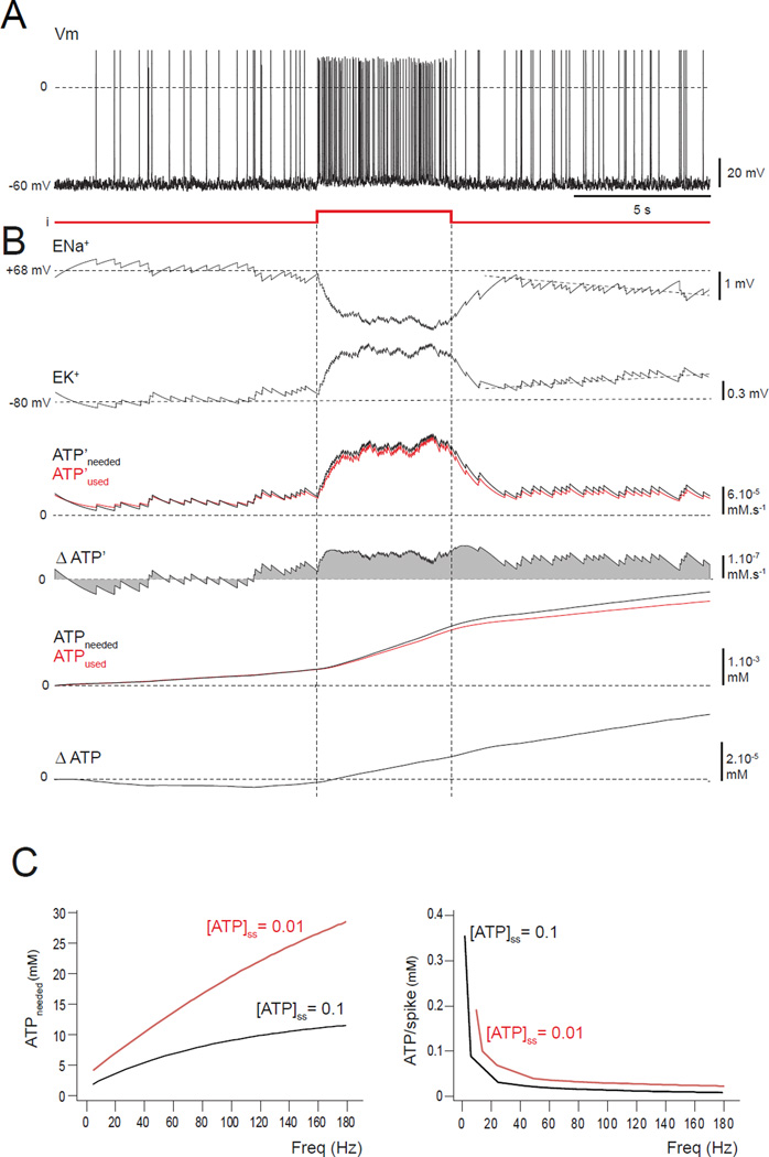
FIGURE 2. A neuron with a small ATP reduction is stable for low-frequency firing but a brief input pulse triggers instability.
(A–C) Model is set with a ten-fold reduction in [ATP]ss, i.e. 0.01 mM.(A) Spiking appears normal before, during and after a brief depolarizing pulse. (B) The reversal potentials ENa+ and EK+ start to drift (tilted dashed lines) from their stable values after the higher-frequency firing, indicating the triggering of an instability. The instability results from the difference between the ATP’used and ATP’needed (red and the black curves) after the pulse, the separation of ATPused and ATPneeded, the positive value of ΔATP’, and the increasing ΔATP. (C) For a total simulated run of 25 s, the amount of ATP needed by the entire neuron was measured for different firing frequencies. When the ATP level is normal ([ATP]ss = 0.1 mM; black curve), the ATP needed (left panel) is lower by a factor of about 3 than with a ten-fold reduction ([ATP]ss = 0.01 mM; red curve). Similar results were found for the ATP needed per AP (right panel). Note that APs are less costly for high-frequency firing.

