Fig. 7.
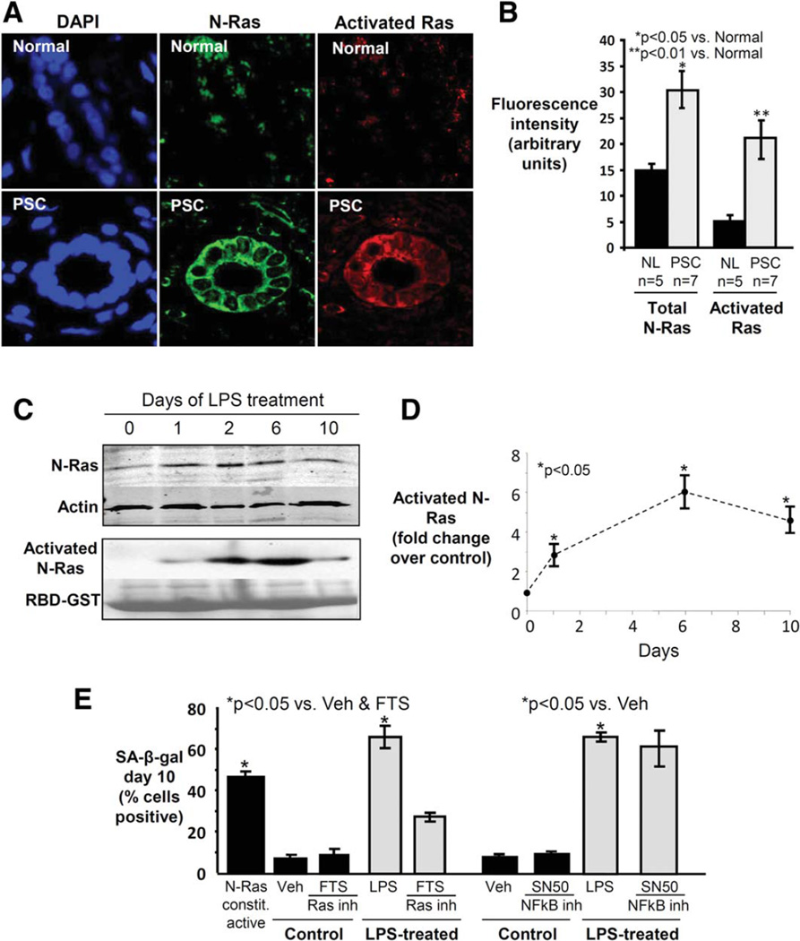
Ras is activated in cholangiocytes in PSC liver, and Ras activation is involved in LPS-induced NHC senescence. (A) Representative con-focal immunofluorescence images for N-Ras (green) and activated Ras (red) (DAPI = blue). Cholangiocyte N-Ras colocalizes with activated Ras in PSC liver, suggesting that N-Ras is not only expressed, but also activated in PSC cholangiocytes. (B) Semiquantitative analysis of fluorescence demonstrates significantly increased N-Ras and activated Ras in PSC cholangiocytes compared to normal. (C) LPS treatment of NHCs induces persistent N-Ras activation. Western blot demonstrates cholangiocyte N-Ras expression, and an RBD-GST pulldown demonstrates LPS-induced Ras activation over the course of 10 days (Ponceau Red used to stain total RBD-GST, loading control). (D) Densitometry on the activated N-Ras blot at each time point. (E) Ras, but not NF-κB inhibition, decreases LPS-induced cholangiocyte senescence. NHCs were cultured in the presence or absence of LPS and a Ras (FTS) or NF-κB (SN50) inhibitor. Cholangiocyte senescence, determined by proportion of SA-β-gal-positive cholangiocytes, is abrogated by Ras inhibition.

