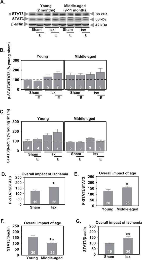
Figure 5.
Effect of age and ischemia on STAT3 in CA1 12 h following global ischemia. Representative Western blots (A) and relative abundance of p-STAT3 and STAT3 (B and C) in CA1 whole-cell lysates from placebo and estradiol (E) treated rats subjected to sham surgery or ischemia (isx). Data are expressed relative to controls (young sham) (B and C). Ischemia increased p-STAT3 and total STAT3 in CA1 of both young and middle-aged rats (D and G; P < 0.05 all ischemic groups vs all sham groups). Middle-aged rats showed higher levels of p-STAT3 than young rats (E; P < 0.05 all middle-aged groups vs all young groups). Estradiol did not alter levels of either p-STAT3 or total STAT3 at 12 h following ischemia (B and C; P > 0.05). Total STAT3 levels were lower in middle-aged than in young rats (F; P < 0.01 all middle-aged groups vs all young groups). *, ** P < 0.05 and P < 0.01, respectively.

