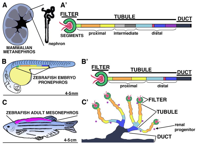
FIGURE 1. Nephron segmental anatomy is conserved among vertebrates.
(A) Schematic of a mammalian metanephros with (A′) a linear diagram of nephron segments. (B) Schematic of a zebrafish embryo with lateral location of the pronephros indicated, and with (B′) a linear diagram of the nephron segments. The zebrafish embryo is approximately 4 mm long (tip to tail) by 5 dpf, and continues to utilize the pronephros as the fish grows. (C) Schematic of a zebrafish adult with dorsal location of the mesonephros, and with (C′) a diagram depicting the arborized arrangements of nephrons with common duct exitways. The adult zebrafish is typically 4–5 cm in length (tip to tail). Analogous nephron segments are color-coded, with the vasculature (red ball), podocytes (dark green), neck (light green), proximal tubule segments (orange, yellow), intermediate tubule segments (gray), distal tubule segments (light blue, dark blue, purple) with intervening macula densa (mammals) or corpuscle of Stannius (fish) (red), and finally the duct (black). [Reprinted from Transl Res, 163(2), McCampbell K, Wingert RA, New Tides: using zebrafish to study renal regeneration, Pages No. 109–122, Copyright 2014, with permission from Elsevier.]

